×

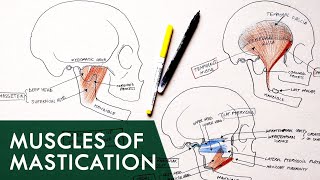
Anatomy Knowledge's video: The Infratemporal Fossa - Boundaries Contents Anatomy Tutorial

@The Infratemporal Fossa - Boundaries & Contents | Anatomy Tutorial
Donation Link: https://paypal.me/studentlamedicina?locale.x=en_US https://www.instagram.com/anatomy.knowledge/ The mandibular nerve exits the skull through foramen ovale. Just under the foramen ovale its gives off a recurrent meningeal branch called the nervus spinosum which enters the skull thorugh foramen spinosum. The otic gg. Is also attached to the mandibular nerve at this level. Soon the mandibular nerve splits into an anterior and a posterior division. From the anterior division are arising branches like the : deep temporal nerves, masseteric nerve and buccal nerve. The posterior division of the mandibular nerves splits into three branches: The inferior alveolar nerve which goes inferiorly to enter the mandibular foramen; The lingual nerve which goes anteroinferiroly; And the auriculotemporal nerve which arises by two roots from the posterior division of the mandibular nerve. The chorda tympani nerve, a branch of the facial nerve, appears in the infratemporal fossa and unites with the lingual nerve. The maxillary artery is one of the two terminal branches of the external carotid artery. The other terminal branch of external carotid is the superficial temporal artery. The maxillary artery arises at a straight angle from the external carotid, passes medial to the neck of the mandibular condyle and goes anteriorly to leave the infratemporal fossa by way of pterygomaxillary fissure. It gives numerous branches like the Middle meningeal artery which goes upwards, passes inbetween the two roots of auriculotemporal nerve and enters the skull by way of foramen spinosum. An accessory meningeal artery may arise to enter the skull by oway of foramen ovale. The inferior alveolar artery follow the course of inferior alveolar nerve; Other branches from the maxillary artery are: The masseteric artery, buccal artery and the anterior and posterior deep temporal arteries. The Pterygoid venous plexus accompanies the maxillary artery and towards the neck of the mandibular condyle gives rise to the maxillary vein. The maxillary vein unites with the superficial temporal vein to form the retromandibular vein. The muscular structures present in the infratemporal fossa are the :Lateral & medial pterygoid muscles , & tendon of temporalis;

1.9K

51
Anatomy Knowledge
Subscribers
131K
Total Post
117
Total Views
1M
Avg. Views
13.4K
View Profile
This video was published on 2020-06-16 23:24:55 GMT by @Anatomy-Knowledge on Youtube. Anatomy Knowledge has total 131K subscribers on Youtube and has a total of 117 video.This video has received 1.9K Likes which are higher than the average likes that Anatomy Knowledge gets . @Anatomy-Knowledge receives an average views of 13.4K per video on Youtube.This video has received 51 comments which are higher than the average comments that Anatomy Knowledge gets . Overall the views for this video was lower than the average for the profile.Anatomy Knowledge #infratemporal #maxillary #mandibular Donation has been used frequently in this Post.

Other post by @Anatomy Knowledge