×

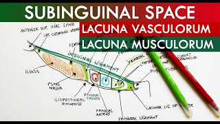
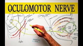
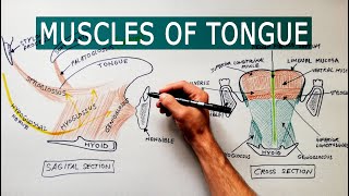
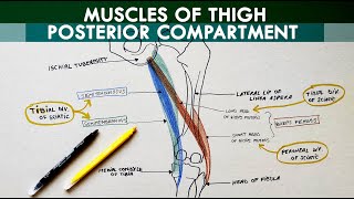
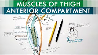
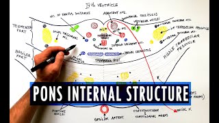
Anatomy Knowledge's video: Anterior view of the brainstem Neuroanatomy Diagram

@Anterior view of the brainstem | Neuroanatomy Diagram
Link for Donations https://paypal.me/studentlamedicina?locale.x=en_US https://www.instagram.com/anatomy.knowledge/ Today we will draw the anatomical features present on the anterior aspect of the brainstem. The bottom part of the brainstem is the medulla which is boundered superiorly by the pontomedullary sulcus and inferiorly by the pyramidal decussation. On the midline we have the anterior median fissure. On each side of this fissure is a proeminence called Pyramid. Lateral to the pyramid is a proeminent oval structure called Olive. Between Pyramid and Olive is the preolivary sulcus. At this level the Hypoglossal nerve or the 12th cranial nerve originates from the medulla. Behind Olive is the retrolivary sulcus. At this level, from above downwards originates the glossopharyngeal nerve or the 9th cranial nerve, The vagus nerve or the 10th cranial nerve and the cranial root of accessory nerve or 11th cranial nerve. The middle portion of the brainstem is represented by the pons which is boundered superiorly by the pontomesencephalic sulcus and inferiorly by the already indicated pontomedullary sulcus. On the midline the pons features a groove for the basilar artery. Laterally , marking the limit between pons and middle cerebellar peduncle is the apparent origin of the trigeminal nerve or the 5th cranial nerve. The trigeminal nerve is comprised of a lateraly large sensory root and a medialy smaller motor root. Beween Pons and Medulla, at the level of Pontomedullary suclus, emerge three cranial nerves. From medial to lateral we have the abducens or the 6th cranial nerve, the facial or 7th cranial nerve and vestobulocochlear or 8th cranial nerve. The top part of the brainstem is the midbrain. The midbrain is boundered superiorly by the optic chiasm and optic tracts. Inferiorly is boundered by the pontomesencephalic sulcus. The ventral surface of the midbrain presents two cerebral peduncles which emerge from the cerebral hemispheres and converge downward to enter the pons. Between cerebral peduncles, optic chiasm and optic tracts is present a space called the interpeducular fossa. At this level from above downward we find the Pituitary stalk, the two mammillary bodies and the posterior perforated substance. The oculomotor nerve or the 3rd cranial nerve is emerging at a groove on the lateral wall of the interpeduncular fossa. On the lateral surface of the cerebral peduncles close to the pontomesencephalic sulcus is the trochlear nerve or the 4th cranial nerve which is the only cranial nerve that exits the brainstem through its posterior surface.

151

12
Anatomy Knowledge
Subscribers
131K
Total Post
117
Total Views
1M
Avg. Views
13.6K
View Profile
This video was published on 2022-10-07 18:14:06 GMT by @Anatomy-Knowledge on Youtube. Anatomy Knowledge has total 131K subscribers on Youtube and has a total of 117 video.This video has received 151 Likes which are lower than the average likes that Anatomy Knowledge gets . @Anatomy-Knowledge receives an average views of 13.6K per video on Youtube.This video has received 12 comments which are lower than the average comments that Anatomy Knowledge gets . Overall the views for this video was lower than the average for the profile.Anatomy Knowledge #brainstem #neuroanatomy #brain has been used frequently in this Post.

Other post by @Anatomy Knowledge