×

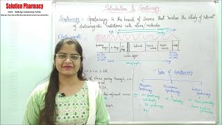
Solution- Pharmacy's video: Anatomy Physiology 96 Structure Function of Eyes Sense Organ Eyes Structure with Functions

@Anatomy & Physiology (96) Structure & Function of Eyes | Sense Organ | Eyes Structure with Functions
Download the "Solution Pharmacy" Mobile App to Get All Uploaded Notes, Model Question Papers, Answer Papers, Online Tests and other GPAT Materials - https://play.google.com/store/apps/details?id=co.bolton.nqilv (01) Eyes are organ of sight located in the deep, protective bony cavities, called the orbits or eye sockets of the skull (02) The eye is about 2.5 cm in diameter (03) The space between the eye and bony orbits is occupied by adipose tissue, connective tissue, ligaments and muscles (04) The bony walls of the orbit and the adipose tissue help protect the eye from injury, and ligament suspends the eye in such a way that the muscle can move it to let one see up, down, and on the side (05) The study of the structure, function and disorder of the eye is termed as “Ophthalmology” Structure of the eye (01) Outer fibrous layer or corneoscleral layer – This layer includes the sclera and cornea (02) Middle vascular layer – Includes – choroid, ciliary body and iris (03) Inner nervous layer – Include retina (01) Sckera - The white of the eye. The sclera gives the eye structural strength and protection for the inner elements of the eye (02) Cornea -This is the outer clear covering over the center of the eye. The cornea acts like both a window and a lens. Light first enters the eye through the cornea. The shape of the cornea then acts as a lens to begin to bend or “refract” light toward a focal point at the back of the eye. The cornea is the primary focusing element of the eye. It is made up of five layers of material. The inner layers add strength and substance to the eye, while the outermost layer provides a tough, protective membrane that helps to shield the more delicate areas of the eye beneath. This outer layer of the cornea is called “epithelium”. Its cells can regenerate in as little as three days. This allows any superficial injury to the outer cornea to heal very quickly. (02) Choroid - The choroid is the posterior portion of the middle layer that lines most of the internal surface of sclera . It contains numerous blood vessels and is composed of soft connective tissue containing pigment cells, melanocytes that produces the melanin pigment. The melanin in choroid is responsible for its dark brown colour. Melanin absorb stray light and darkens the eyeball cavity, which prevents the internal reflection and scattering of light within the eyeball. As a result the image remain sharp and clear (03) Retina - This is the focusing screen of the eye. The retina is made up of nerve tissue. This keenly sensitive nerve tissue lines the rear wall of the eye. The retina receives the finely focused light from the lens and turns the light energy into electrical impulses. These impulses can then be translated into images by the brain. The retina processes light through a layer of photoreceptor cells. These are essentially light-sensitive cells, responsible for detecting qualities such as color and light-intensity. The retina processes the information gathered by the photoreceptor cells and sends this information to the brain via the optic nerve. Basically, the retina processes a picture from the focused light, and the brain is left to decide what the picture is. Due to the retina’s vital role in vision, damage to it can cause permanent blindness. Conditions such as retinal detachment, where the retina is abnormally detached from its usual position, can prevent the retina from receiving or processing light. This prevents the brain from receiving this information, thus leading to blindness. Get in touch with the solution by just clicking the following links- Facebook Group- https://www.facebook.com/groups/solutionpharamcy Mobile App - https://play.google.com/store/apps/details?id=co.bolton.nqilv New Channel (Pharmacy Dictionary) https://www.youtube.com/channel/UCt6OXVV_2oxf5DD0Mad6e9A E-Mail for official and other work - solutionpharmacy@gmail.com

24

2
Solution- Pharmacy
Subscribers
0.9M
Total Post
2.5K
Total Views
393.9K
Avg. Views
2.3K
View Profile
This video was published on 2024-02-07 21:00:08 GMT by @Solution--Pharmacy on Youtube. Solution- Pharmacy has total 0.9M subscribers on Youtube and has a total of 2.5K video.This video has received 24 Likes which are lower than the average likes that Solution- Pharmacy gets . @Solution--Pharmacy receives an average views of 2.3K per video on Youtube.This video has received 2 comments which are lower than the average comments that Solution- Pharmacy gets . Overall the views for this video was lower than the average for the profile.Solution- Pharmacy #solutionpharmacy #Pharmacologyclass #Pharmacognosyvideos #GPAT has been used frequently in this Post.

Other post by @Solution Pharmacy